This page is available in other languages:
EIZO productcatalogus
Met behulp van door EIZO zelf ontwikkelde software kunt u snel en eenvoudig de eigenschappen en instellingen van uw eigen monitoren testen.
Met de ColorNavigator-software kunt u ColorEdge-beeldschermen eenvoudig en nauwkeurig kalibreren.
Met de EIZO-software kunt u het gebruik en de configuratie voor een configuratie met één scherm of meerdere monitoren eenvoudig beheren. Windows- en macOS-besturingssystemen worden ondersteund.
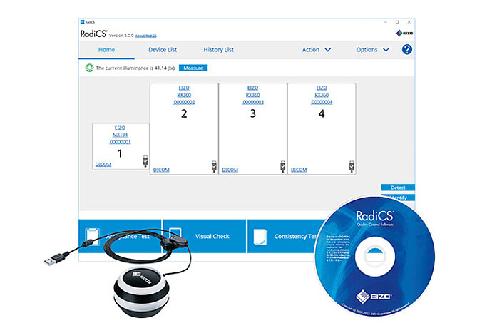
Met de software van EIZO kunt u het volledige kwaliteitsbeheerproces aansturen - van de kalibratie en het asset-management tot aan de overdrachts- en consistentiecontrole.
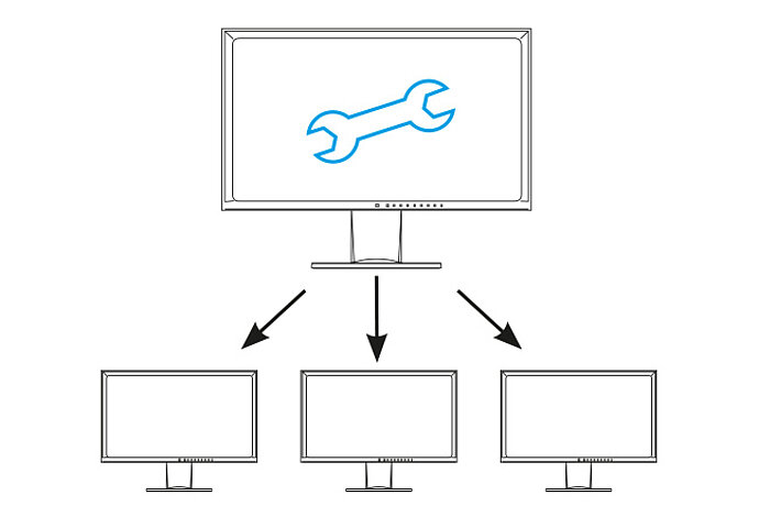
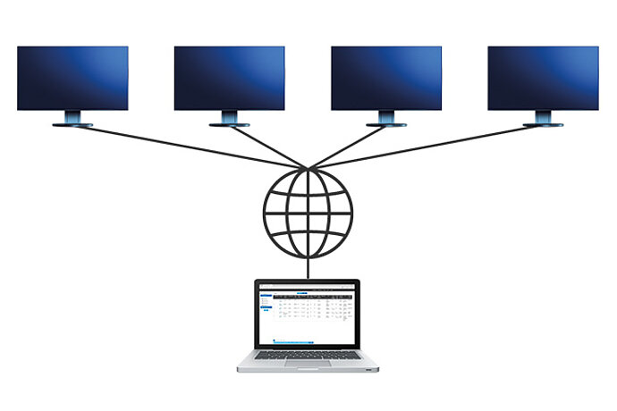
EIZO Monitor Configurator vergemakkelijkt netwerkoverschrijdende installatie als monitoren in een bedrijf dezelfde instellingen moeten hebben.
Met Quick Color Match-software wordt het complexe kleurenbeheerproces voor het afdrukken met inkjetprinters voor thuisgebruik heel eenvoudig.
RadiCS LE is kwaliteitsmanagementsoftware waarmee u EIZO RadiForce-monitoren kunt kalibreren en kalibratiegegevens kunt beheren.
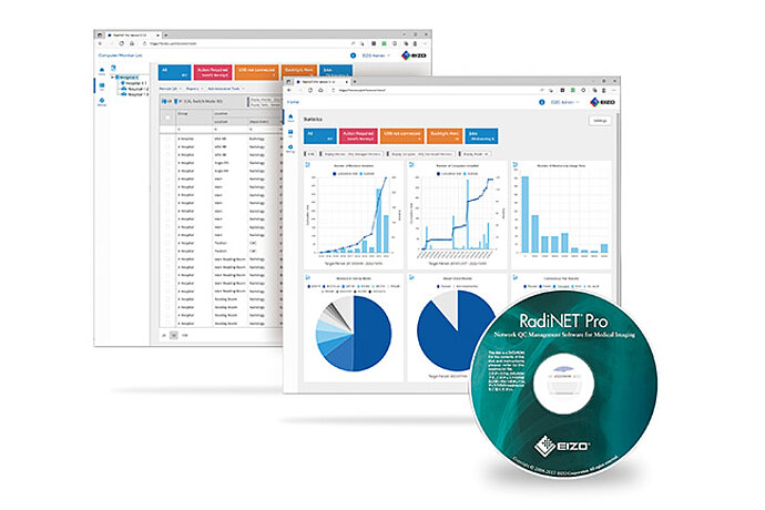
Speciale software van EIZO voor netwerkondersteund kwaliteitsbeheer in grote installaties. Met functionaliteit voor externe bediening van monitoren.
ColorNavigator Network maakt de centrale beeldkwaliteitsborging van ColorEdge-monitoren mogelijk.
Met de servertoepassing Screen InStyle Server kunnen systeembeheerders alle monitoren en PC’s in een netwerk instellen en beheren.
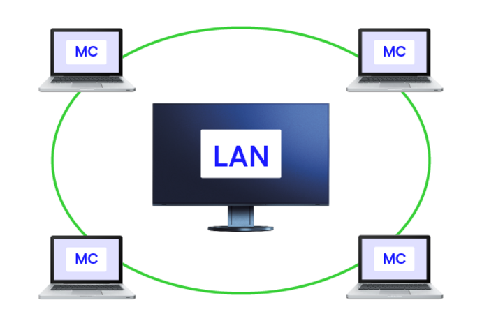
Software voor het tijdelijk vervangen van het MAC-adres van een EIZO monitor met LAN-aansluiting door het geverifieerde MAC-adres van een aangesloten PC.
11 van 11 producten worden weergegeven